


- Refer a Patient
- Referral Types
- Patient Information
- Overview of CFEH Clinics
- CFEH Instrument List
- Our clinical team
- Causes of Vision Loss
- Patient Forms


An extract from the CFEH Atlas
Choroideraemia
Overview
Choroideraemia is an X-linked condition affecting the RPE, outer retina, and choriocapillaris. It is caused by mutations in the CHM gene. Due to this inheritance pattern, choroideraemia is much more common in males than in females.
The disease process is characterised by nyctalopia and peripheral vision loss in the second decade of life or before. In the early stages, peripheral pigmentary mottling may be noted and over time this develops into pigment clumping and atrophy of the RPE, photoreceptors, choriocapillaris and eventually, choroidal atrophy follows.
Over time, loss of the RPE means that the choroidal vasculature becomes more visible. As the disease progresses and further atrophy of the choroid occurs (usually in the mid-peripheral and peripapillary fundus first) and the bare sclera may become visible – seen clinically as areas of pale white areas on retinal imaging.
Pigment clumping occurs as a result of RPE degeneration. This pigment can be seen on OCT as hyper-reflective deposits. Also seen on OCT are interlaminar bridges (ILBs) which are hypo-reflective wedge-shaped structures that bridge the inner and outer retina in choroideraemia. Outer retinal tubulations are seen in late-stage disease on OCT. These appear as hyper-reflective round structures with a hypo-reflective lumen. The tubulations are composed of deteriorating photoreceptors and remnants of the external limiting membrane.
In recent studies, OCT angiography has been used to further our knowledge around choroideraemia. These studies showed that the choriocapillaris shows greater nonperfusion than the retina in choroideraemia.
Disease progression is relatively slow with central vision being fairly well preserved until late stage disease starts to affect the macula – typically around 50 to 70 years of age.
Being an x-linked condition, females are rarely diagnosed with choroideraemia, however the carriers of the defective gene may show a partial expression of the condition. Most carriers are asymptomatic and visual acuity is unaffected, however the fundus may show signs consistent with choroideraemia such as pigment mottling.
- Case 1: Choroideraemia
- Case 2: Advanced disease
- Case 3: Carrier
A 42-year-old caucasian male with best-corrected visual acuity of 6/9.5 (20/30) in the right eye and 6/12 (20/40) in the left.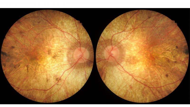
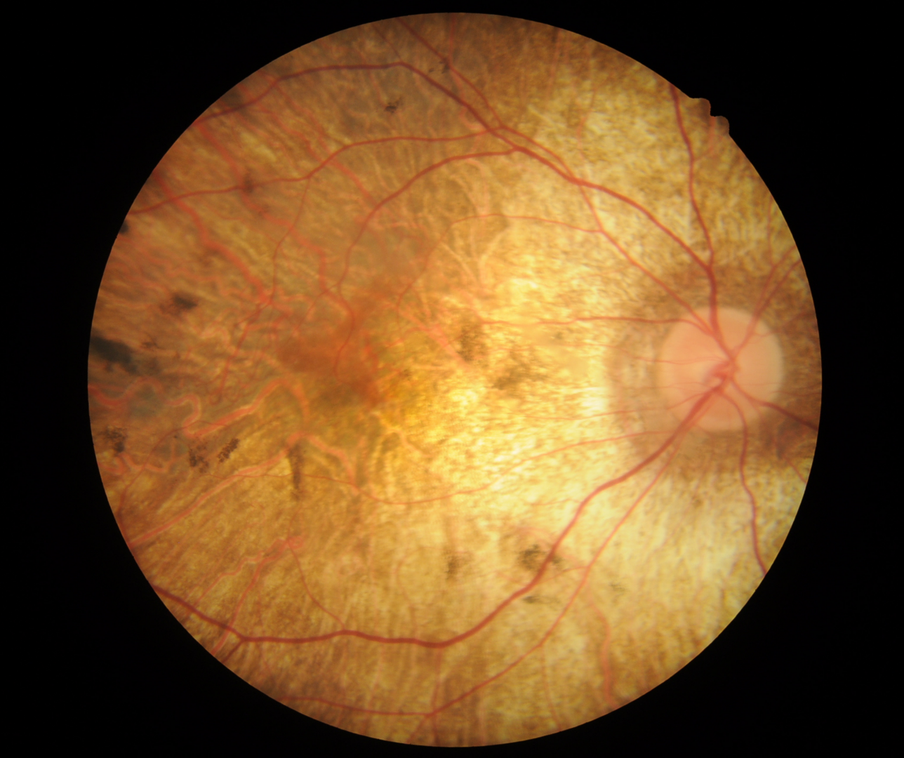
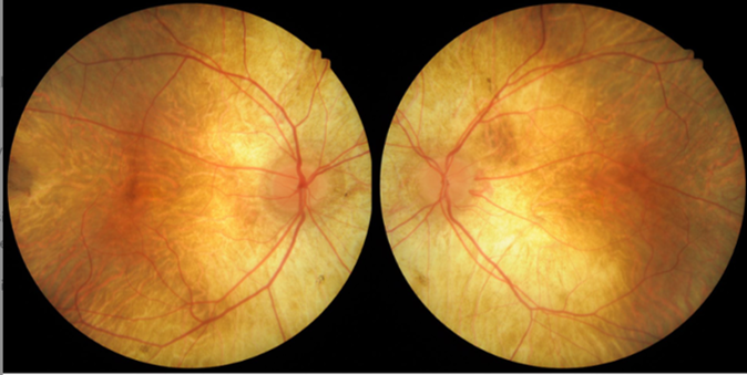
Posterior pole photos (right and left eye): Colour fundus photographs show atrophy of the macula and peripapillary areas and scattered pigment clumps.
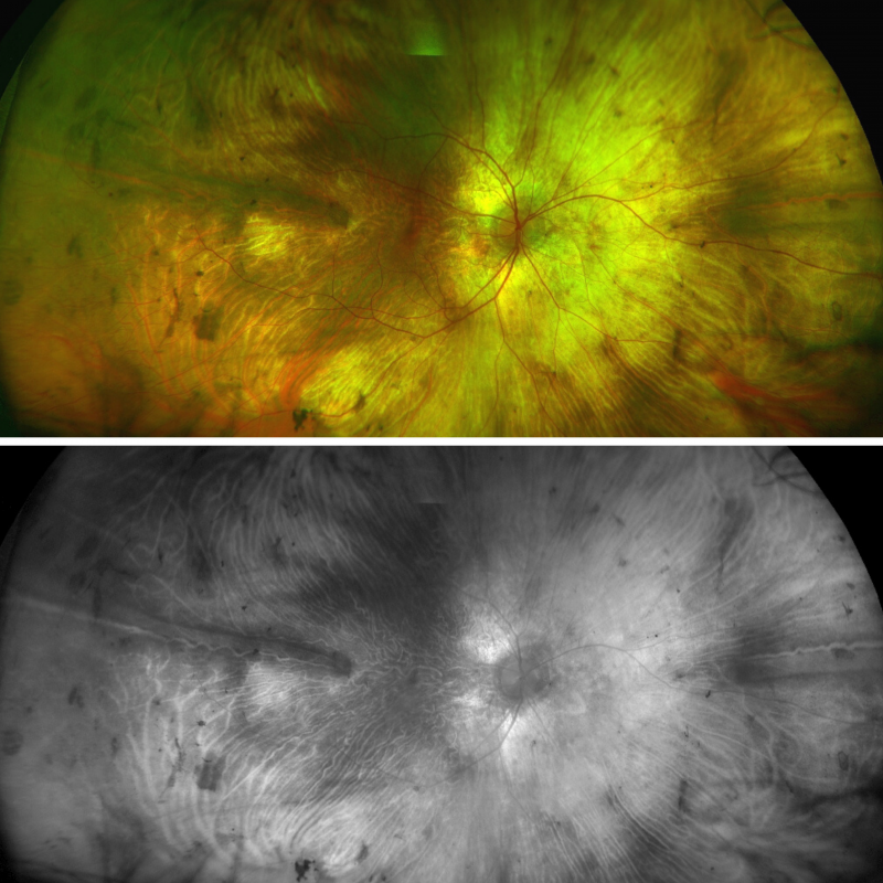
Optomap Widefield Images (right and left eye): Widefield imaging shows more pigment clumps in the midperipheral retina. The choroidal vessels in the mid-periphery and peripheral retina show increased visibility due to widespread loss of the RPE throughout the retina.
Optomap Widefield Green-Separation Images (Right and Left eye): These images highlight the clumps of pigment scattered throughout the retina.
Fundus Autofluorescence (FAF) Images: Fundus autofluorescence shows a relatively normal autofluorescent small islands of the central macula and peripapillary area with hypo-autofluorescent areas beyond.
OCT Spectralis line and volume scan (right eye): OCT shows extensive loss of the photoreceptors, RPE, choriocapillaris and choroid and outer retinal tubulations adjacent to a small island of remaining functioning retina.
OCT Spectralis line and volume scan (left eye): OCT shows extensive loss of the photoreceptors, RPE, choriocapillaris and choroid and outer retinal tubulations adjacent to a small island of remaining functioning retina.
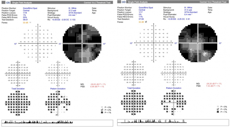
10-2 SITA Standard Visual Field: Central 10-2 visual fields show significant loss of the central visual field.
A 72-year-old Caucasian male with best-corrected visual acuity of 6/12+ (20/40+) and 6/38 (6/127) in the left eye. He reports longstanding night vision difficulties and poor peripheral vision.
Posterior pole photos (right and left eye): Colour fundus photographs show diffuse chorioretinal atrophy at the posterior pole.
Optomap and green separation images (right eye): Widefield imaging shows increased choroidal vessel visibility, large areas of visible sclera and pigment clumping in the mid-periphery and peripheral retina.
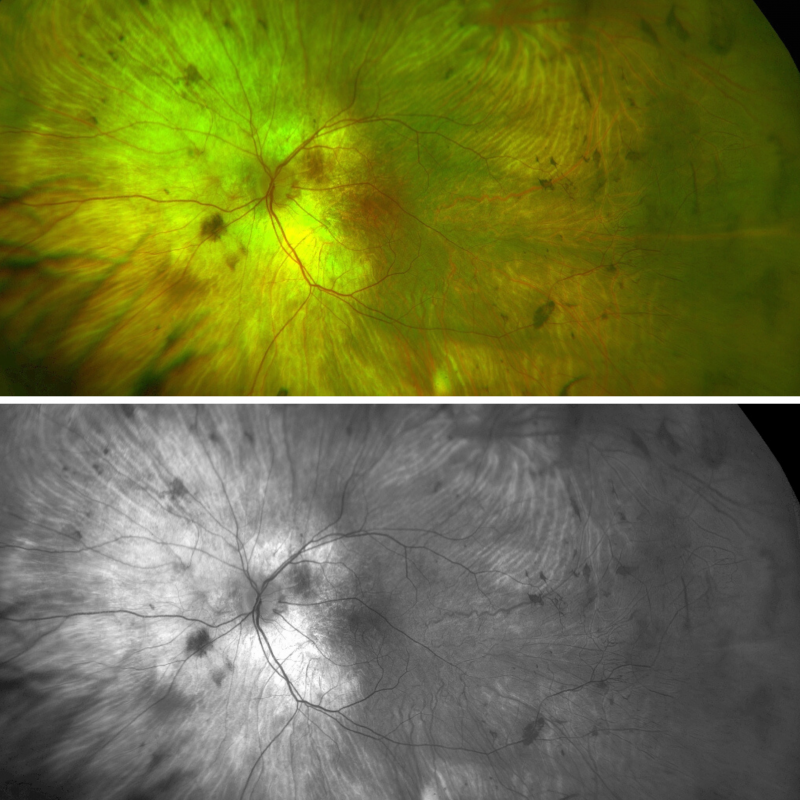
Optomap and green separation images (left eye): Widefield imaging shows increased choroidal vessel visibility, large areas of visible sclera and pigment clumping in the mid-periphery and peripheral retina.
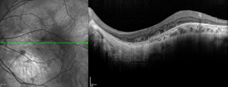
Spectralis OCT line scan (left macula): OCT shows outer retinal tubulations. There is a complete loss of the RPE and choroid in the nasal half of this image.
A 55-year-old female with best-corrected visual acuity of 6/6 (20/20) in each eye. The patient’s father had choroideraemia and was legally blind at age 40. She reports no difficulty with night vision.
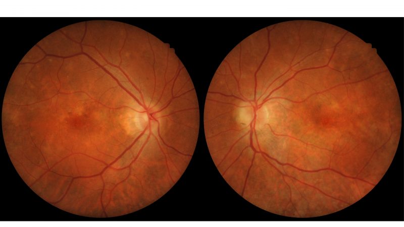
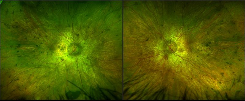
Colour fundus photographs (right and left eye): The retina has a mottled appearance with pigmentary changes apparent at both maculae.
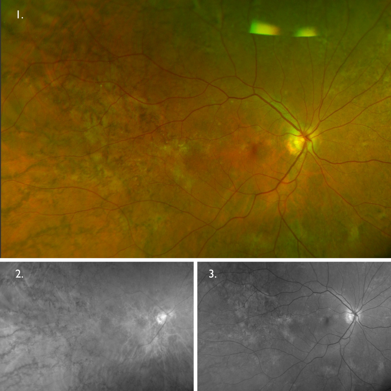
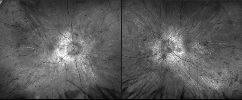
Optomap (1), red separation (2) and green separation (3) images – right eye: Widefield imaging shows reticular-type pigmentation throughout the retina, being more dense peripherally.
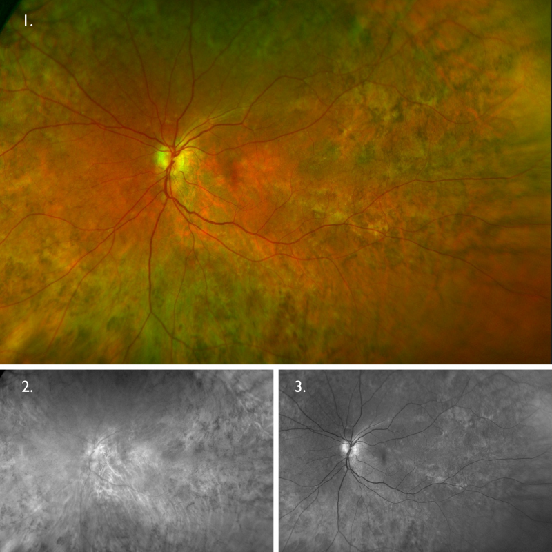
Optomap (1), red separation (2) and green separation (3) images – left eye: Widefield imaging shows reticular-type pigmentation throughout the retina, being more dense peripherally. These changes are slightly more extensive in the left eye than they are in the right.
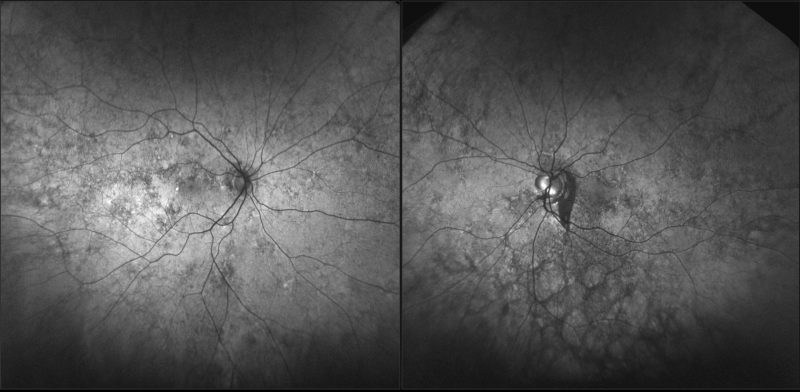
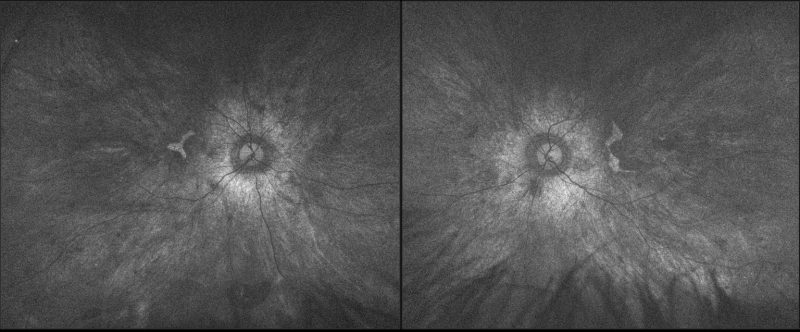
Fundus autofluorescence imaging (right and left eye): Fundus autofluorescence imaging shows several hyperfluorescent spots in the central retina of both eyes, as well as hypofluorescent patches across the posterior pole and periphery corresponding to the reticular pigmentary changes in each eye. There is an area of hypofluorescence temporal to the optic nerve in the left eye extending inferiorly.
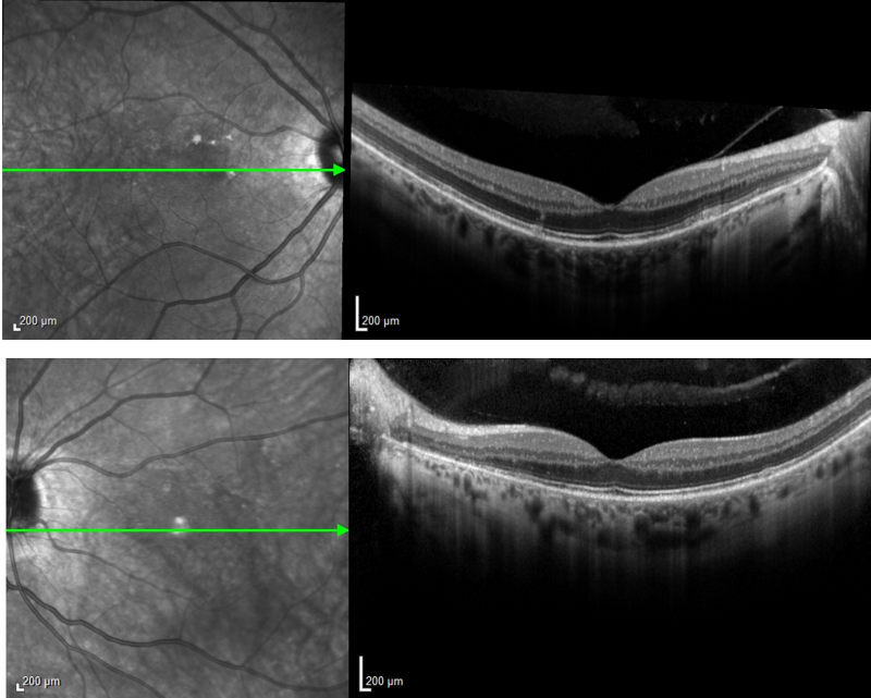
Spectralis macular OCT scans show essentially normal anatomy at both maculae with few small elevations of the RPE
Differential Diagnosis
References
MacDonald, IM. Russell, L. Chan, CC (2009) Choroideremia: New Findings from Ocular Pathology and Review of Recent Literature, Survey of Ophthalmology, Volume 54, Issue 3, Pages 401-407.
Sun, LW, Johnson, RD, Williams, V. Summerfelt, P. Dubra, A. Weinberg, DV, Stepien, KE, Fishman, GA, Carroll, J. (2016). Multimodal Imaging of Photoreceptor Structure in Choroideremia. PLoS One, 11(12)
Zinkernagel, MS. & MacLaren, RE. (2015). Recent advances and future prospects in choroideremia. Clinical Ophthalmology, 9, 2195-2200.