(02) 8115 0700
Centre for Eye Health is an initiative of Guide Dogs NSW/ACT and the University of NSW.
Our work is possible thanks to the generosity of Guide Dogs NSW/ACT and their supporters.
To support us, please donate.

CFEH Instrument List
Kowa Wx3D Non-mydriatic Retinal Camera (Kensington)
Captures en face colour fundus images with capabilities of stereoscopic viewing of the optic nerve head
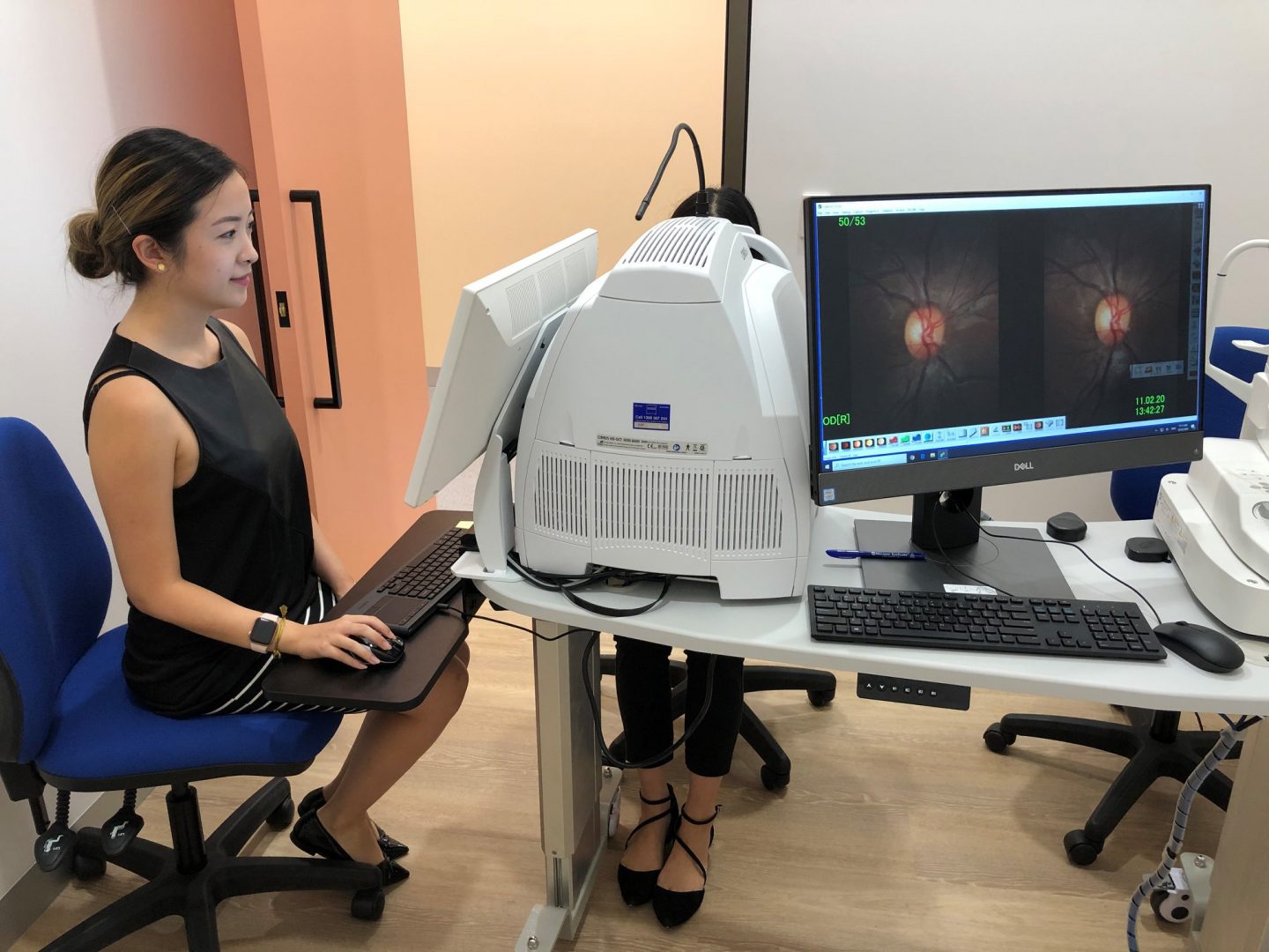
Cirrus Spectral Domain HD-OCT including OCT Angiography
Produces high resolution in vivo cross sectional images of the posterior eye allowing measurement of RNFL, macula and ganglion cell thickness as well as glaucoma progression analyisis, OCT angiography and anterior segment assessment.
Spectralis OCT including OCT Angiography and anterior OCT
Produces high resolution in vivo cross sectional images of the posterior eye allowing measurement of RNFL, macula and ganglion cell thickness as well as OCT angiography, anterior segment assessment and fundus autofluorescence imaging
Optomap Wide-Field Fundus Photography
Wide-field fundus photography.
Heidelberg HRT3
Produces optic nerve head morphological images (glaucoma module) allowing topographical change assessment over time and can also take en face confocal microscopy images of the cornea (rostock cornea module)
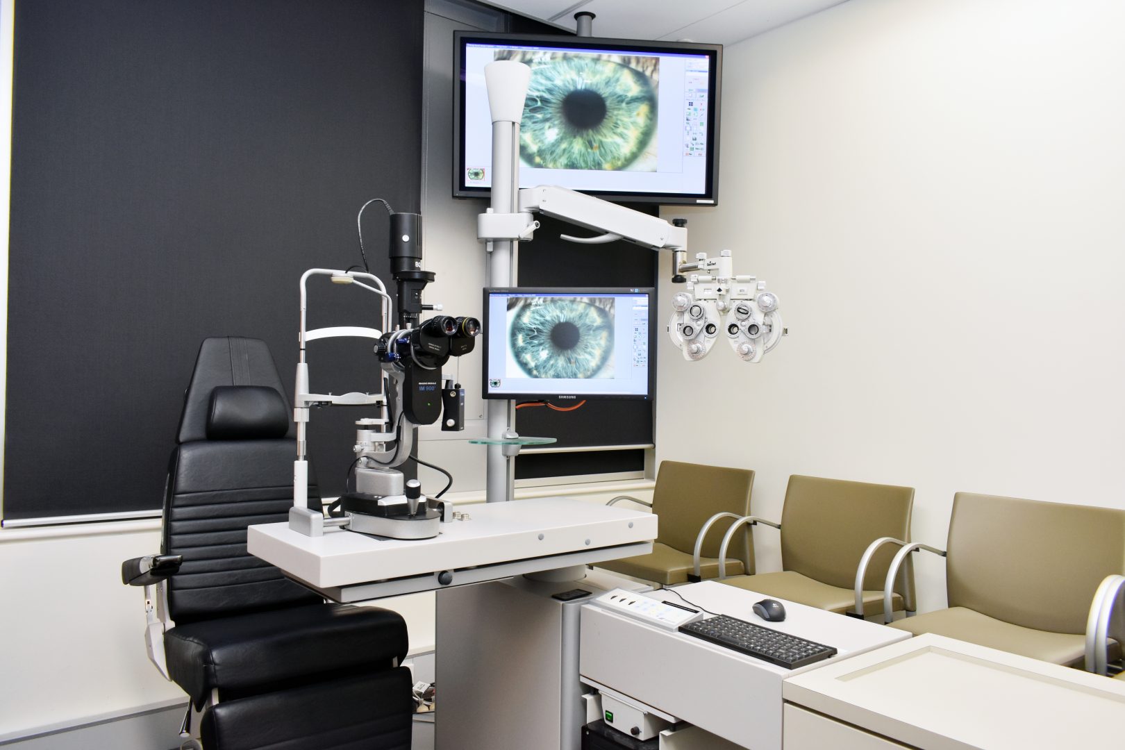
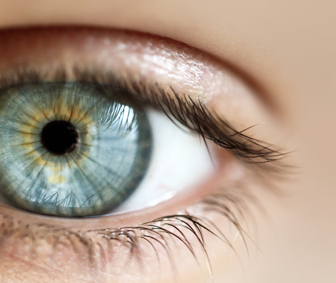
Haag-Streit Video and Photography Slit Lamp Imaging
Video and still digital imaging of the anterior segment to document anterior segment pathology.
Nidek Confoscan 4
En face confocal microscopy of the cornea with up to 500x magnification. Uses include measuring epithelial/total corneal thickness and endothelial cell count.
Lensstar - Haag-Streit Optical Biometry Using Partial Coherence Interferometry (Sutherland)
Measures axial length, corneal thickness, anterior chamber depth, crystalline lens thickness, vitreous chamber depth, retinal thickness and calculates IOL power. Used in assessment for cataract surgery, glaucoma assessment and monitoring myopia progression.
Humphrey Visual Field Analysis
Measures sensitivity of the visual field using white-on-white or blue/yellow stimuli plus glaucoma progression analysis
Humphrey Matrix Perimetry
Measures sensitivity of the visual field using frequency-doubling stimuli.
iCare Tonometer and iCare Home
Intraocular pressure measurement without corneal anaesthesia. iCare home allows diurnal phasing of IOP to detect fluctuations in measurements.
Pachmate Ultrasonic Pachymetry (DGH pachmate)
Measures total corneal thickness
ImagineEyes Wavefront Aberrometry (IRX3)
Measures total eye refractive aberrations, modulation transfer and point spread function and Strehl ratio in preparation for cataract or refractive surgery and in cases of keratoconus.
OCULUS Keratograph 5M
A corneal topographer with built-in real keratometer and a color camera optimized for external imaging. Used for examining meibomian glands, non-invasive tear film break-up time, measuring tear meniscus height and evaluating the lipid layer.
OCULUS Pentacam Scheimpflug Photography
Measures front and back surface topography, anterior chamber depth and volume, total corneal pachymetry, corneal aberration analysis and crystalline lens densitometry
Tomey A-Scan and B-Scan Ultrasonography
A-Scan biometry and B-Scan eye cross-section/morphology.
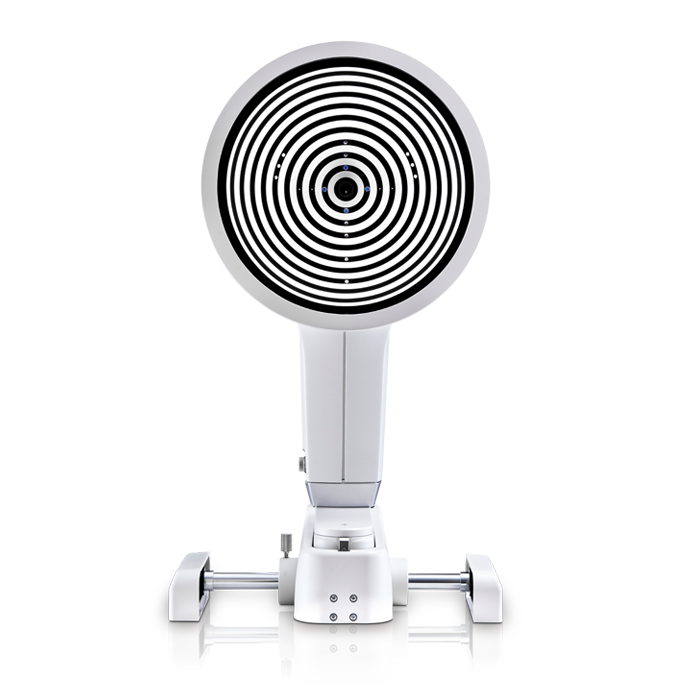
Medmont Corneal Topography (E300)
Measures corneal topography and curvature variations across the corneal surface.
(02) 8115 0700
Centre for Eye Health is an initiative of Guide Dogs NSW/ACT and the University of NSW.
Our work is possible thanks to the generosity of Guide Dogs NSW/ACT and their supporters.
To support us, please donate.