Extract from the CFEH Atlas
Ocular Albinism
Overview
Albinism is an inherited condition involving an abnormality in melanin pigment production or impairment of the maturation of melanosomes. Oculocutaneous albinism affects the eyes, skin, and hair and is an autosomal recessive disorder. Ocular albinism affects the eyes only and typically shows an X-linked inheritance pattern. It is caused by mutations in GPR143 gene.
Ocular albinism is characterised by reduced visual acuity, nystagmus, various grades of iris translucency, fundus hypopigmentation and foveal hypoplasia. Foveal hypoplasia is caused by the underdevelopment of the foveal pit and has been associated with poor visual acuity. The foveal pit and the foveal avascular zone are either reduced or absent. Another characteristic sign is chiasmal misrouting, which refers to an increased number of retinal ganglion cells from the temporal retina projecting to the contralateral hemisphere and can be detected by visually evoked potential (VEP).
Female carriers of X-linked ocular albinism type can show partial iris translucency and scattered areas of depigmentation but are otherwise asymptomatic.
Case Examples
- Case 1
- Case 2
- Case 3
A 21-year-old Caucasian female with best-corrected acuities of 6/7.5 (20/25) in each eye.
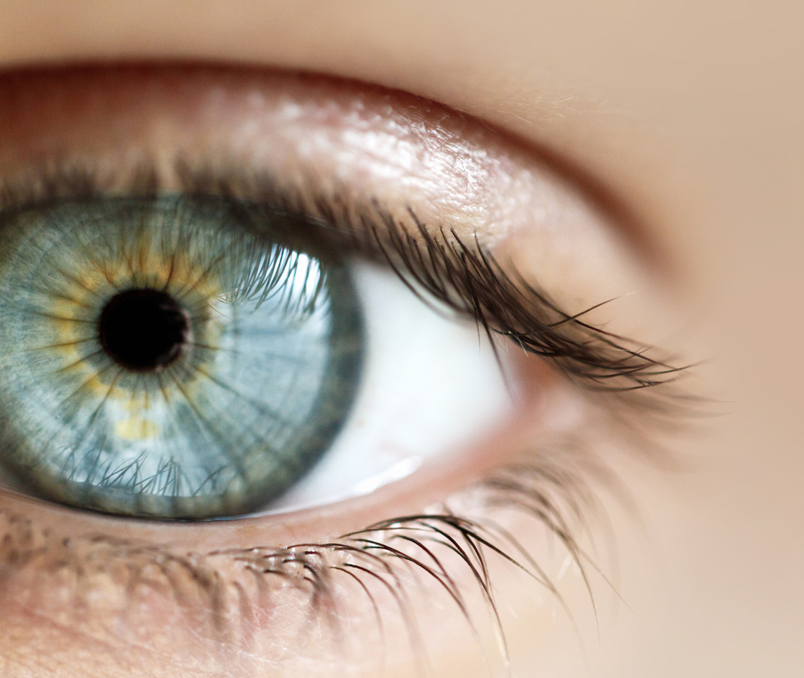
Anterior eye photo (right eye): Anterior photograph shows punctuate iris translucency.
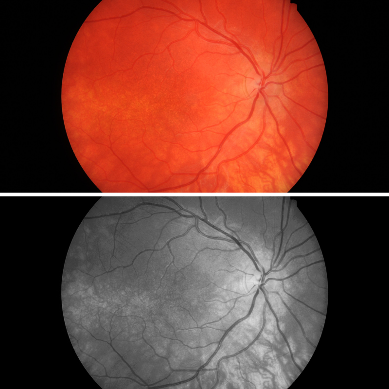
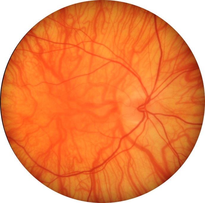
Fundus photographs (and red-free images) show a poorly defined foveal area and mottling of the macular pigmentation. Choroidal vessels are visible in the posterior pole but not in the macula (grade 2 fundus hypopigmentation).
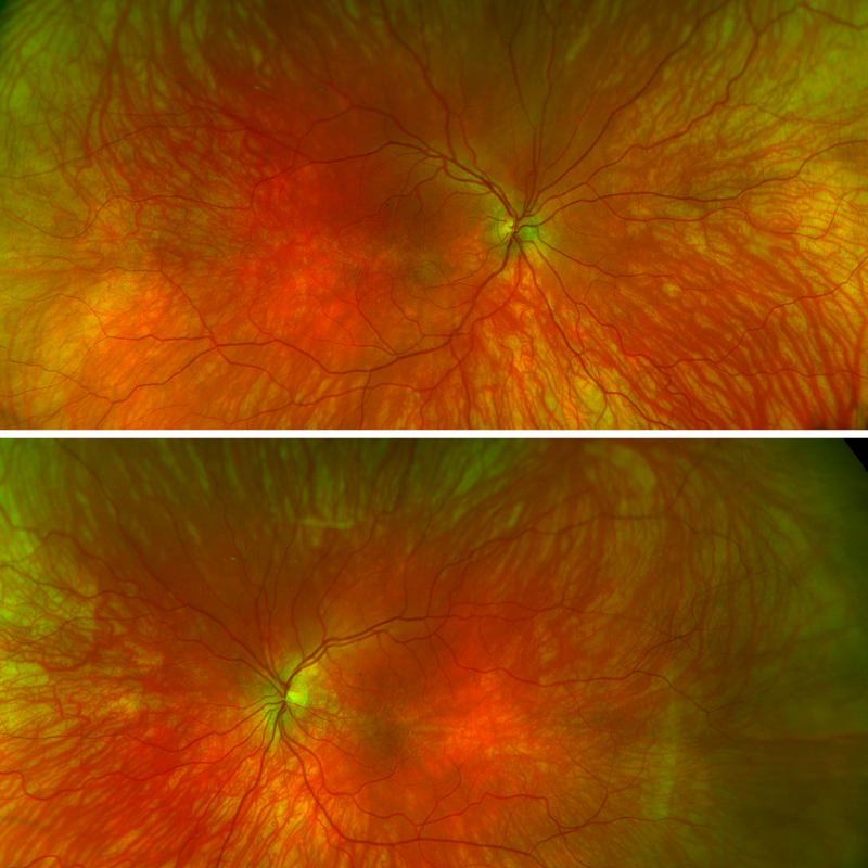
Optomap images (right and left eye): Widefield imaging displays the fundus hypopigmentation. The choroidal vessels are highly visible, particularly in the midperipheral retina.
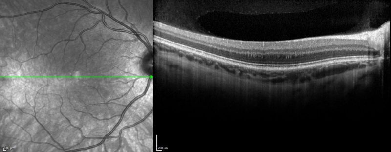
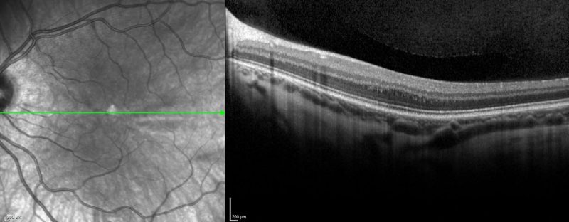
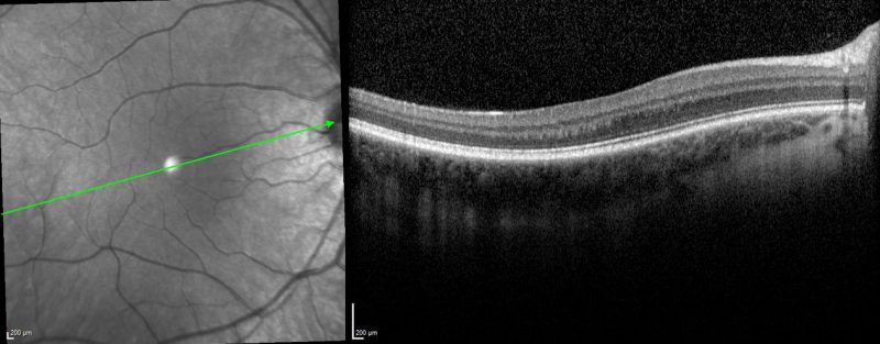
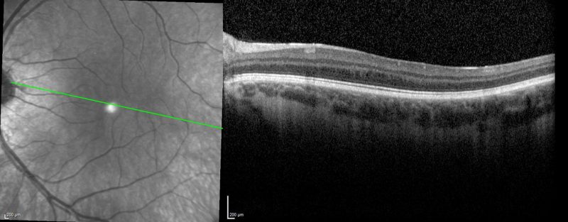
Spectralis OCT line scan (right and left macula): OCT imaging shows the absence of the normal anatomical foveal depression.
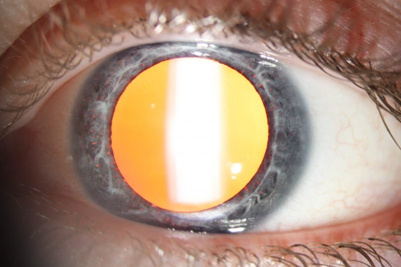
A 47-year-old female with nystagmus and best-corrected acuities of 6/75 (20/250) in the right eye and 6/60 (20/200) in the left. Slit-lamp examination revealed marked iris transillumination defects.
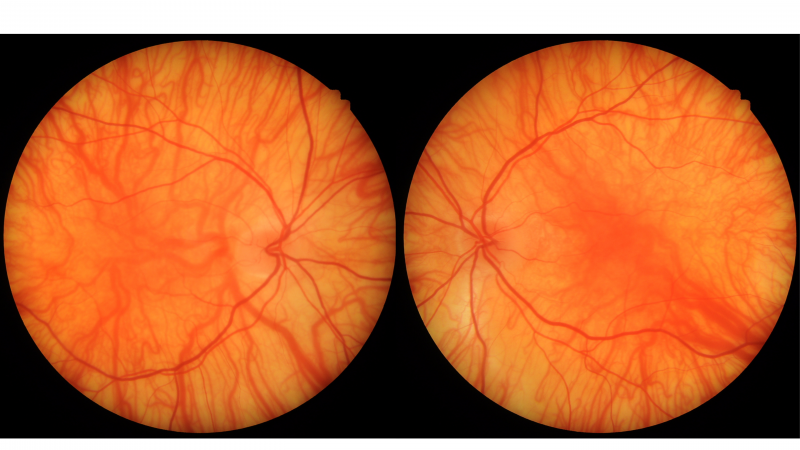
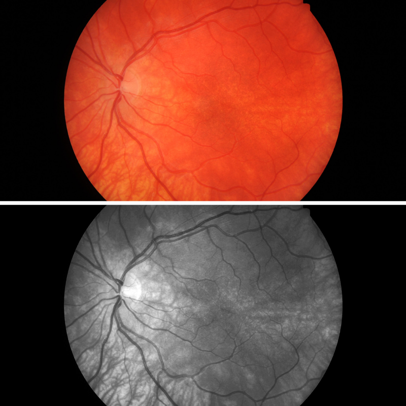
Fundus photographs show grade 3 fundus hypopigmentation with highly visible choroidal vasculature in the macula region. The fovea is poorly defined.
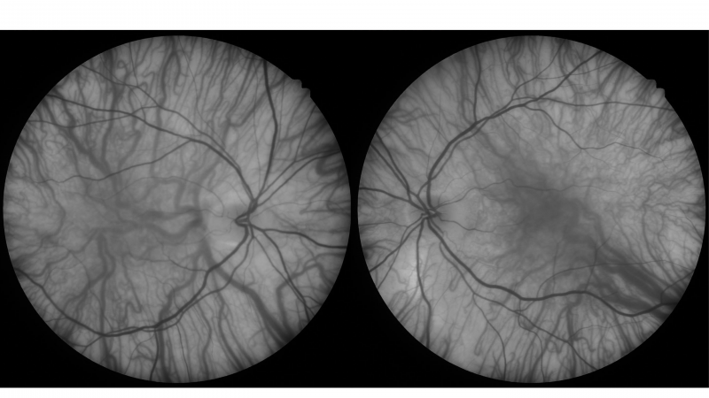
Red-free images (right and left eye): Red-free images highlight the lack of retinal pigmentation and increased visibility of the choroid.
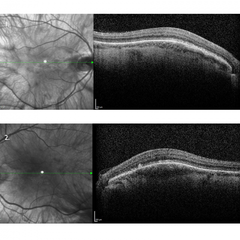
Spectralis OCT line scans (right and left macula): OCT macular line scans show the absence of a foveal pit with no widening of outer nuclear layer in both eyes. This is grade 4 foveal hypoplasia and is associated with poor visual acuity.
A 26-year-old male with best-corrected visual acuity of 6/7.5 (20/25) in each eye.
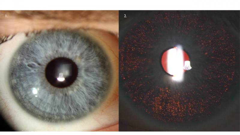
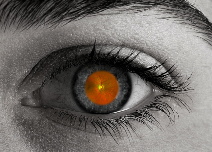
Anterior eye photo (1) and retroillumination (2): The anterior photograph shows diffuse iris translucency.
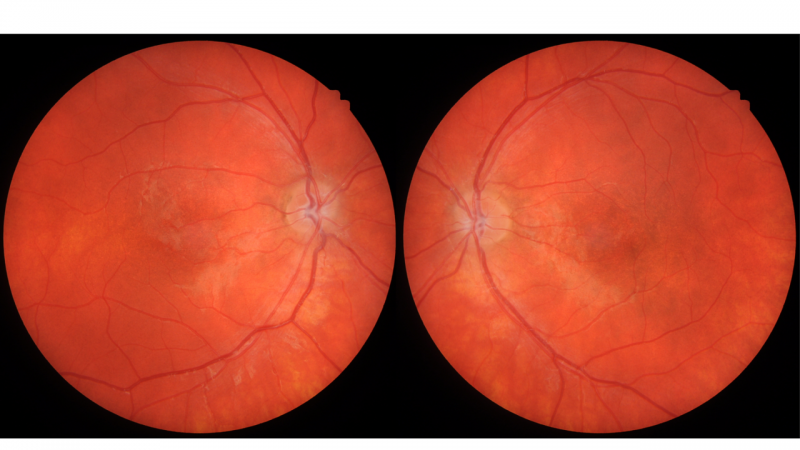
Fundus photography (right and left): Colour fundus photographs show mottled pigmentation at both maculae with the fovea poorly defined. Choroidal vessels are not visible in the posterior pole.
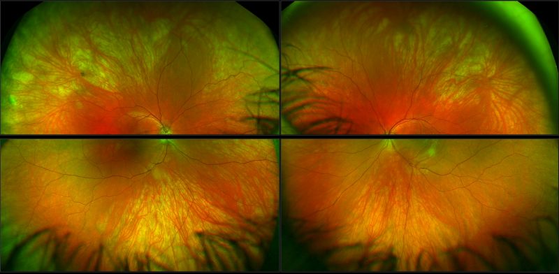
Optomap images (superior and inferior retina, right and left eyes): Widefield imaging shows grade 1 fundus hypopigmentation with visible choroidal vessels in the midperiphery.
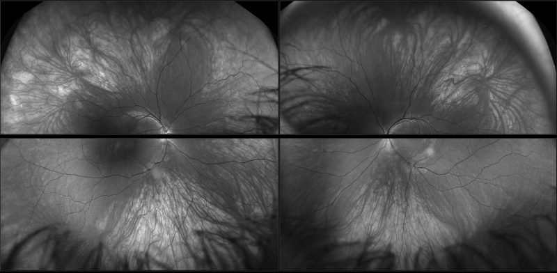
Green separation images (superior and inferior retina, right and left eyes): These images highlight the lack of retinal pigmentation and increased visibility of the choroid.
Spectralis OCT volume and line scans (right and left macula): OCT line reveals absence of the foveal pit.
Differential diagnosis
Another differential to consider is diffuse myopic chorioretinal atrophy. More information about this condition can be found in the CFEH Atlas.
Foveal Hypoplasia
Foveal hypoplasia can also occur in several conditions including achromatopsia, aniridia, microphthalmos, retinopathy of prematurity, Incontinentia pigmenti and Stickler syndrome and can also be an isolated idiopathic condition.
References
Kruijt CC, de Wit GC, Bergen AA et al. The Phenotypic Spectrum of Albinism. Ophthalmology 2018; 125: 1953-1960.
Thomas MG, Kumar A, Mohammad S et al. Structural Grading of Foveal Hypoplasia Using Spectral-Domain Optical Coherence Tomography: A Predictor of Visual Acuity? Ophthalmology 2011; 118: 1653-1660.
Apkarian P, Reits D, Spekreijse H et al. A decisive electrophysiological test for human albinism. Electroencephalography and Clinical Neurophysiology 1983; 55: 513-531.
Kondo H. Foveal hypoplasia and optical coherence tomographic imaging. Taiwan J Ophthalmol 2018; 8: 181-188.